The medial pterygoid (or internal pterygoid muscle), is a thick, quadrilateral muscle of mastication.

The mandibular branch of the fifth cranial nerve, the trigeminal nerve, innervates the medial pterygoid muscle.
Structure
It consists of two heads.
- The bulk of the muscle arises as a deep head from just above the medial surface of the lateral pterygoid plate.
- The smaller, superficial head originates from the maxillary tuberosity and the pyramidal process of the palatine bone.
Its fibers pass downward, lateral, and posterior, and are inserted, by a strong tendinous lamina, into the lower and back part of the medial surface of the ramus and angle of the mandible, as high as the mandibular foramen. The insertion joins the masseter muscle to form a common tendinous sling which allows the medial pterygoid and masseter to be powerful elevators of the jaw.
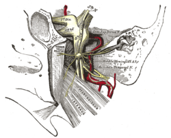
Innervation
Medial pterygoid is innervated by nerve to medial pterygoid (a branch of the mandibular nerve), which also innervates tensor tympani and tensor veli palatini.
Unlike the lateral pterygoid and all other muscles of mastication which are innervated by the anterior division of the mandibular branch of the trigeminal nerve, the medial pterygoid is innervated by the main trunk of the mandibular branch of the trigeminal nerve (V), before the division.
Function
Given that the origin is on the medial side of the lateral pterygoid plate and the insertion is from the internal surface of the ramus of the mandible down to the angle of the mandible, its functions include:
- Elevation of the mandible (closes the jaw)
- Minor contribution to protrusion of the mandible
- Assistance in mastication
- Excursion of the mandible; contralateral excursion occurs with unilateral contraction.
This article uses material from the Wikipedia article
Metasyntactic variable, which is released under the
Creative Commons
Attribution-ShareAlike 3.0 Unported License.
Metasyntactic variable, which is released under the
Creative Commons
Attribution-ShareAlike 3.0 Unported License.