The peroneus brevis muscle (or fibularis brevis muscle) lies under cover of the peroneus longus, and is the shorter and smaller of the peroneus muscles.

Structure

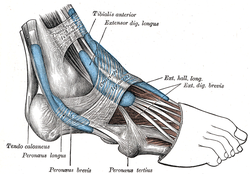
It arises from the lower two-thirds of the lateral surface of the body of the fibula, medial to the peroneus longus, and from the intermuscular septa separating it from the adjacent muscles on the front and back of the leg.
The fibers pass vertically downward, and end in a tendon which runs behind the lateral malleolus along with but in front of that of the preceding muscle, the two tendons being enclosed in the same compartment and lubricated by a common mucous sheath.
It then runs forward on the lateral side of the calcaneus, above the calcaneal tubercle and the tendon of the peroneus longus, and is inserted into the tuberosity at the base of the fifth metatarsal bone, on its lateral side. When the base of the fifth metatarsal is fractured, the peroneus brevis may pull on and displace the proximal fragment (Jones Fracture). An inversion sprain of the foot may pull the tendon such that it avulses the tuberosity at the base of the fifth metatarsal.
Nerve supply
It is innervated by the superficial fibular nerve, also known as the superficial peroneal nerve.[1]
Function
The peroneus brevis muscle is the strongest abductor of the foot.[2] It also assists in weak plantarflexion and eversion of the foot.[2] It provides lateral stability to the foot and ankle.[3]
History
Etymology
The terms "Peroneal" (i.e., Artery, Retinaculum) and "Peroneus" (i.e., Longus and Brevis) are derived from the Greek word Perone (pronounced Pair-uh-knee) meaning pin of a brooch or a buckle. In medical terminology, both terms refer to being of or relating to the fibula or to the outer portion of the leg.
Additional images
| This article uses material from the Wikipedia article Metasyntactic variable, which is released under the Creative Commons Attribution-ShareAlike 3.0 Unported License. |